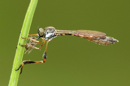
Leptogastrinae sensu Dikow 2009a

Beameromyia disfascia , image © Michael Thomas.
Leptogastrinae
– abdominal T2 more than twice as long as wide (153)
– S2 divided medially into two equal halves, which are separated by fenestra (159)
– seta-like spicules on hypopharynx spaced far apart (33)
– ocellar setae absent (62)
– only regular setae form katatergal setae (83)
– presutural dorsocentral setae absent (92)
– lateral depression on prothoracic coxae absent (104)
– row of macrosetae on anterodorsal mesothoracic tibiae absent (110)
– proximal prothoracic and mesothoracic tarsomeres longer than following two tarsomeres combined (120, 121)
– pulvilli absent (123)
– claws fairly straight throughout (125)
– R4 and R5 more or less parallel (133)
– R4 terminating posterior to wing apex (143)
– female spermathecal reservoir formed by more or less expanded ducts to sac-shaped reservoir (180)
– female spermathecal reservoir heavily sclerotized (181)
– male lateral ejaculatory process present, large cylindrical sclerite (213)
The monophyly of Leptogastrinae has never been in question and is corroborated by many character states. Hull (1962) included the genus Acronyches within Leptogastrinae, but this placement was not acknowledged by Martin (1968). As Acronyches is the sister taxon to the Leptogastrinae sensu previous authors and two autapomorphies characterise this clade, Dikow (2009) includes this genus in the newly delimited Leptogastrinae.
Acronychini: Acronyches
– vertex sharply depressed (3)
– frons markedly approximating medially at level of antennal insertion (48)
– metkatepisternum large and visible between mesothoracic and metathoracic tibiae (100)
– setiform empodium minute or entirely absent (127)
– microtrichia on posterior margin of wing arranged in two divergent planes (141)
– female S8 plate-like and hypogynial valves separated (surrounded by membrane) (170)
– female furca divided into two lateral sclerites (184)
Leptogastrini: Beameromyia, Euscelidia, Lasiocnemus, Leptogaster, Tipulogaster
– postpronotal lobes extended medially and anteriorly and nearly touching medially (72)
– metathoracic coxae directed anteriorly (111)
– male with surstylus on epandrium (198)
– male with lateral process of gonostyli present (207)
– only lower facial margin developed (4)
– dorsoposterior margin of cibarium with one transverse ridge connecting cornua (35)
– cornua on cibarium well developed in anteroposterior orientation (37)
– postpedicel tapering distally (54)
– postpronotum setae absent (70)
– anterior anepisternal and proepimeral setae erect to appressed and anteriorly directed (78, 79)
– postmetacoxal bridge partly present laterally and membranous area medially (102)
– cell cup open (136)
– female with short spermathecal ducts (178)
– male with gonocoxite or gonocoxite-hypandrial complex partially fused to epandrium (203)
– male lacking gonocoxal apodemes (204)
– male with sperm sac appearing more or less heavily sclerotized (218)
